
File:Validation of the dye diffusion assay performed with the flattened cochlear preparation.png

預覽大小:287 × 599 像素。 其他解析度:115 × 240 像素 | 230 × 480 像素 | 979 × 2,044 像素。
原始檔案 (979 × 2,044 像素,檔案大小:3.33 MB,MIME 類型:image/png)
摘要
| 描述 |
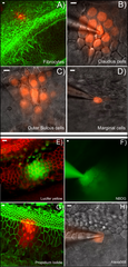
English: A–D: Dye diffusion patterns after PI was injected into a single cell in various locations in the cochlea. The type of the cells that was injected is given at lower right corner of each panel. E–F: Diffusion patterns of four different fluorescent dyes after injecting into a single Claudius cell. Name of the dye is given in the lower right corner of each panel. Panels B), C), D), F) & H) were photographed with unfixed fresh samples. Panels A), E), G) were results obtained from fixed samples after the experiments were done. They were labeled with fluorescent phalloidin (red in E, green in A&G) to outline the cell border. Scale bar on the top left of each panel represents approximately 100 µm. |
| 日期 | |
| 來源 | PLOS ONE an open source peer reviewed journal- Gap Junction Mediated Intercellular Metabolite Transfer in the Cochlea Is Compromised in Connexin30 Null Mice ([1]) |
| 作者 | Qing Chang, Wenxue Tang, Shoeb Ahmad1, Binfei Zhou1, Xi Lin1 |
授權條款
此檔案採用創用CC 姓名標示 2.5 通用版授權條款。
- 您可以自由:
- 分享 – 複製、發佈和傳播本作品
- 重新修改 – 創作演繹作品
- 惟需遵照下列條件:
- 姓名標示 – 您必須指名出正確的製作者,和提供授權條款的連結,以及表示是否有對內容上做出變更。您可以用任何合理的方式來行動,但不得以任何方式表明授權條款是對您許可或是由您所使用。
檔案歷史
點選日期/時間以檢視該時間的檔案版本。
| 日期/時間 | 縮圖 | 尺寸 | 使用者 | 備註 | |
|---|---|---|---|---|---|
| 目前 | 2009年2月12日 (四) 04:24 | | 979 × 2,044(3.33 MB) | Mike.lifeguard | {{Information |Description={{en|1=A–D: Dye diffusion patterns after PI was injected into a single cell in various locations in the cochlea. The type of the cells that was injected is given at lower right corner of each panel. E–F: Diffusion patterns o |
檔案用途
下列頁面有用到此檔案:
全域檔案使用狀況
以下其他 wiki 使用了這個檔案:
- en.wikipedia.org 的使用狀況
- pt.wikipedia.org 的使用狀況